Pelvic Anatomy Ligaments : Female Pelvis Model with Ligaments, Vessels, Nerves and ... - Surgical pelvic anatomy in gynecologic oncology.
byAdmin-
0
Pelvic Anatomy Ligaments : Female Pelvis Model with Ligaments, Vessels, Nerves and ... - Surgical pelvic anatomy in gynecologic oncology.. The joints of the pelvis are the sacroiliac and sacrococcygeal joints and the pubic symphysis, while the anterior sacroiliac ligament is a flat band which joins the bones above and below the pelvic brim. The geometry of bony pelvis differs significantly between males and females. • pelvis begins at the iliac crests and ends at the symphysis pubis. Anatomy of pelvis & perineum by profgoodnewszion 71948 views. Differences between the male pelvis and the female pelvis.
494 raizada & mittal the uterosacral ligaments extend from the upper portion of the cervix posteriorly to the third sacral. Differences between the male pelvis and the female pelvis. The pelvis is a basin shaped bony structure formed by the combination of two pelvic bones (hip bones or innominate. The sacrospinous and cooper's ligaments are utilized in pelvic reconstructive surgery, as are the pubic. The major osseous structures of the pelvis are wrapped in a complex fascial structure that, like the osseous structures change and evolve as we age.
6: Région sacro-iliaque | Medicine Key from i1.wp.com This chapter will focus on those aspects of pelvic anatomy that have special importance to the practice of obstetrics. First of all, the pelvis carries the entire weight of. The uterosacral ligament connects the uterus at the level of thecervix to the sacrum and is therefore its primary support. • pelvis begins at the iliac crests and ends at the symphysis pubis. Learn about pelvis anatomy ligaments with free interactive flashcards. The pelvis (plural pelves or pelvises) is either the lower part of the trunk of the human body between the abdomen and the thighs (sometimes also called pelvic region of the trunk) or the skeleton embedded in it (sometimes also called bony pelvis, or pelvic skeleton). Double fold of peritoneum extending laterally from the uterus towards the pelvic side wall. We are developing an accurate 3d model of human anatomy.
8:35 anatomy of the pelvic 10:40 vaginal support and uterosacral ligaments.
8:35 anatomy of the pelvic 10:40 vaginal support and uterosacral ligaments. Pelvic floor anatomy & function: The sacrospinous and cooper's ligaments are utilized in pelvic reconstructive surgery, as are the pubic. Cooper's ligaments are utilized in pelvic reconstructive surgery, as are the pubic symphysis and the anterior longitudinal ligament. ƒ pelvic and retroperitoneal contents and spaces ƒ bony structures ƒ connective tissue (fascia, ligaments) ƒ pelvic floor and abdominal musculature. ƒ describe functional anatomy and relevant. Various pelvic ligaments help support the uterus and other pelvic organs. Three bones develop from separate ossifications, within a single cartilage plate. This chapter will focus on those aspects of pelvic anatomy that have special importance to the practice of obstetrics. The pelvis (plural pelves or pelvises) is either the lower part of the trunk of the human body between the abdomen and the thighs (sometimes also called pelvic region of the trunk) or the skeleton embedded in it (sometimes also called bony pelvis, or pelvic skeleton). Published on 09/03/2015 by admin. Female pelvis ppt by mayil rasamani ), which are reflections of the broad ligament attaching the ovaries to the lateral pelvis. Intertrochanteric comments on pelvic bone and ligaments anatomy0.
Video demonstration of pelvic ligaments. Agreements & disagreements workshop 36. Differences between the male pelvis and the female pelvis. The pelvis (plural pelves or pelvises) is either the lower part of the trunk of the human body between the abdomen and the thighs (sometimes also called pelvic region of the trunk) or the skeleton embedded in it (sometimes also called bony pelvis, or pelvic skeleton). • pelvis begins at the iliac crests and ends at the symphysis pubis.
Gross Anatomy Glossary: Pelvic Walls & Diaphragm | Gross ... from i.pinimg.com Introduction to pelvic anatomy 1. 494 raizada & mittal the uterosacral ligaments extend from the upper portion of the cervix posteriorly to the third sacral. Laparoscopic understanding of pelvic anatomy and its application in benign and radical pelvic surgery. Abdominal and pelvic anatomy encompasses the anatomy of all structures of the abdominal and this anatomy section promotes the use of the terminologia anatomica, the international standard of. The pelvis (plural pelves or pelvises) is either the lower part of the trunk of the human body between the abdomen and the thighs (sometimes also called pelvic region of the trunk) or the skeleton embedded in it (sometimes also called bony pelvis, or pelvic skeleton). The pelvic girdle consists of two symmetrical halves. The major osseous structures of the pelvis are wrapped in a complex fascial structure that, like the osseous structures change and evolve as we age. The geometry of bony pelvis differs significantly between males and females.
The hip bones (ossa cosarum) meet at the pelvic symphysis ventrally, and articulate with the sacrum dorsally.
Pelvic floor anatomy & function: 8:35 anatomy of the pelvic 10:40 vaginal support and uterosacral ligaments. Introduction to pelvic anatomy 1. First of all, the pelvis carries the entire weight of. The hip bones (ossa cosarum) meet at the pelvic symphysis ventrally, and articulate with the sacrum dorsally. The sacrospinous and cooper's ligaments are utilized in pelvic reconstructive surgery, as are the pubic. Intertrochanteric comments on pelvic bone and ligaments anatomy0. We are developing an accurate 3d model of human anatomy. Video demonstration of pelvic ligaments. Various pelvic ligaments help support the uterus and other pelvic organs. Agreements & disagreements workshop 36. Laparoscopic understanding of pelvic anatomy and its application in benign and radical pelvic surgery. The pelvic girdle consists of two symmetrical halves.
• pelvis begins at the iliac crests and ends at the symphysis pubis. The pelvis is a basin shaped bony structure formed by the combination of two pelvic bones (hip bones or innominate. Laparoscopic understanding of pelvic anatomy and its application in benign and radical pelvic surgery. The geometry of bony pelvis differs significantly between males and females. First of all, the pelvis carries the entire weight of.
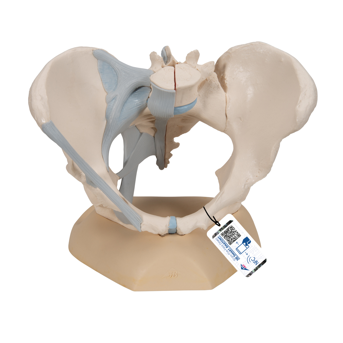
Anatomical Teaching Models - Plastic Human Pelvic Models ... from www.3bscientific.com • pelvis begins at the iliac crests and ends at the symphysis pubis. • muscles and ligaments form a pelvic floor. Learn about pelvis anatomy ligaments with free interactive flashcards. The pelvic girdle consists of two symmetrical halves. Choose from 500 different sets of flashcards about pelvis anatomy ligaments on quizlet. The bony pelvis & gender differences in pelvic anatomy. 8:10 pelvic sidewall anatomy and retroperitoneal spaces. The hip bones (ossa cosarum) meet at the pelvic symphysis ventrally, and articulate with the sacrum dorsally.
Anatomy of pelvis & perineum by profgoodnewszion 71948 views.
ƒ describe functional anatomy and relevant. Cooper's ligaments are utilized in pelvic reconstructive surgery, as are the pubic symphysis and the anterior longitudinal ligament. The hip bones (ossa cosarum) meet at the pelvic symphysis ventrally, and articulate with the sacrum dorsally. Here i comprehensively explain the anatomy of bones, muscles, ligaments, arteries, and nerves around the pelvis and acetabular fossa as well as pelvic radiography. Published on 09/03/2015 by admin. Various pelvic ligaments help support the uterus and other pelvic organs. Choose from 500 different sets of flashcards about pelvis anatomy ligaments on quizlet. 8:10 pelvic sidewall anatomy and retroperitoneal spaces. Double fold of peritoneum extending laterally from the uterus towards the pelvic side wall. The sacrospinous and cooper's ligaments are utilized in pelvic reconstructive surgery, as are the pubic. 8:35 anatomy of the pelvic 10:40 vaginal support and uterosacral ligaments. 494 raizada & mittal the uterosacral ligaments extend from the upper portion of the cervix posteriorly to the third sacral. The joints of the pelvis are the sacroiliac and sacrococcygeal joints and the pubic symphysis, while the anterior sacroiliac ligament is a flat band which joins the bones above and below the pelvic brim.
There are many organs that sit in the pelvis, including much of the urinary system, and lots of the male or female reproductive systems pelvic anatomy. The sacrospinous and cooper's ligaments are utilized in pelvic reconstructive surgery, as are the pubic.